Chn
Chn veta-grand@mail.ru
  +7 499 250 40 00

The Ministry of Health and Social Development of the Russian Federation approved and allowed for wide use in practical health care the following medical technology using the photosensitizer "FOTODITAZIN®" in traumatology:
PHOTODYNAMIC THERAPY OF INFLAMMATORY JOINT DISEASES IN CHILDREN AND ADOLESCENTS
- Juvenile chronic arthritis
- Juvenile rheumatoid arthritis
At the moment, in several medical institutions in Russia, an experimental and clinical assessment of the effectiveness of the use of photodynamic therapy with the drug "Photoditazin®" in traumatology and orthopedics is being carried out.
In the course of the work, methods of photodynamic therapy with the drug "Photoditazin®" (gel penetrator of light radiation) for injuries and degenerative-dystrophic lesions of the knee, shoulder and wrist joints were developed and improved.
Videofluorescent arthroscopic navigation in photodynamic therapy of pathologies of large joints.
Research objectives. In this work, the accumulation of a photosensitizer in large human joints before, during and after a session of photodynamic therapy was studied using new methods and new diagnostic equipment.
Materials and methods. An apparatus and a method have been developed that make it possible to carry out fluorescent video navigation in the inflamed tissues of the human shoulder and knee joints previously photosensitized using the Photoditazin preparation. The equipment is unique in its operating modes; the system is capable of simultaneously displaying two images on the screen: color and fluorescent at the same time.
The concentration of the photosensitizer is determined at any desired point in the image digitally in the monitoring mode. Fluorescence was excited by a laser light source with a wavelength of 635 nm. The control of the therapeutic dose of laser irradiation was monitored by photobleaching in the red wavelength range, which made it possible to carry out diagnostics even in the presence of thin layers of blood on the tissues under study. The method readings were checked using a fiber-optic spectrometer.
Results and Conclusions. The method has demonstrated sensitivity to the contrast of the photosensitizer fluorescence intensity in normal and inflamed joint tissues, which, in turn, makes it possible to draw a conclusion about the contrast of the photosensitizer accumulation and the possibility of using photodynamic therapy. Also, the method has proven its applicability for monitoring the course of photodynamic therapy by the degree of photosensitizer burnout.
Arthroscopic photodynamic therapy for pigmented-villous synovitis of the knee joint
Stranadko E.F1., Ivannikov S.V2., Zharova T.A2., Gear N.A2. Shesternya N.А2.
(1- Federal State Budgetary Institution "State Research Center for Laser Medicine, FMBA of Russia",
2- 2- First Moscow State Medical University named after I.M.Sechenov)
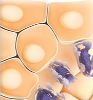
Pigment-villous synovitis is a type of inflammatory process in the synovial membrane. It is manifested by the proliferation of synovial villi, which look red-brown with yellow patches of pigment, their significant compaction due to the accumulation of xanthoma cells, along with fibroblasts. In addition, there is a local disorder of lipid metabolism.
There is an increase in local temperature associated with pronounced vascularization of synovial villi. Lymphocytes, histiocytes, neutrophils, synovial and degenerating cells are found in the synovial fluid.
Growing dense villi begin to fill the cavity of the knee joint and interfere with movements, can cause blockages, which disrupts the biomechanics of the knee joint. Infringement of the synovial villi and their pressure on the surrounding tissues also causes pain in the knee joint area, hemarthrosis. All this disrupts the function of the knee joint and causes the development of deforming arthrosis.
The main method of treatment is the total removal of the pathologically altered synovium. This is done by arthroscopic or arthrotomy excision of the altered villi - their mechanical removal from the joint [21,23-25].
In the future, in postoperative treatment, patients are prescribed courses of non-steroidal anti-inflammatory drugs and are dynamically observed.
In case of relapse, repeated removal of villi is inevitable. The disadvantage of this method is that the method does not stop the development of synovitis, it progresses, repeated surgical interventions on the knee joint are required, which injures and destroys the knee joint.
Among other methods of treatment, radiation therapy is known. It is often used in combination with surgical excision of the villi [20,22,26]. A positive effect is noted, but complications are possible due to radiation exposure to the patient.
We believe it is expedient to use photodynamic therapy (PDT) for arthroscopic debridement of the knee joint.
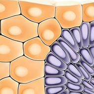
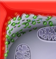
PDT is a loco-regional organ-preserving method for the treatment of various diseases, both malignant tumors and a number of non-neoplastic diseases. The essence of the method is that a photosensitizer, which has a unique property of selectively accumulating in tissues with increased metabolism, is activated by local irradiation of pathologically altered tissues with light with a wavelength corresponding to the long-wave absorption peak of the photosensitizer. The photochemical reaction arising in the presence of oxygen from the tissues themselves causes the generation of singlet oxygen and other reactive oxygen species, which have a detrimental effect on tumor cells, which leads to tumor resorption, or on other pathological tissues with increased metabolism, for example, rheumatoid.
The action of reactive oxygen species is manifested by direct cytotoxic damage to tumor cells or by destruction of blood vessels feeding the tumor or other inflamed tissues. The direct cytotoxic effect of PDT leads to apoptosis of pathologically altered cells, and the vascular mechanism of PDT action leads to hemorrhagic necrosis of the tumor, the vascular network of which differs from the blood supply normal and inflamed tissue. In addition, a whole cascade of immunological reactions develops in the process of PDT, and the products of tumor degradation under the influence of PDT, according to experimental data, act as a specific antitumor vaccine. There is every reason to believe that products by the action of PDT of inflamed tissues also stimulate immune mechanisms. The vascular mechanism of PDT action accounts for about 60% of the therapeutic effect of PDT, direct cytotoxic damage to pathologically altered tissues - about 30%, and the immune mechanisms - a little more than 10%.
It should be noted that in recent years more and more attention has been paid to the immune mechanisms, and their role in photodynamic damage to tumors and other pathological tissues is recognized more and more significant. This is especially true for inflammatory processes.
PDT opens up wide opportunities for various therapeutic effects of a radical and palliative nature, when other methods of treatment have already exhausted themselves or are not applicable at all.
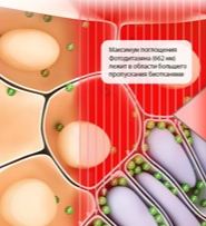
In our work, we used a second-generation photosensitizer from the group of chlorine derivatives, photoditazine, and low-energy laser radiation of a semiconductor laser with an exciting light wavelength of 662 nm.
Chlorins are cyclic tetrapyrroles (porphyrins with a hydrogenated D ring). These are preparations of natural origin, they are obtained from the biomass of the microalga Spirulina platensis - Spirulina platensis. The compositions used in clinical practice contain chlorin eb (70-97%) as the main component.
Compared to first-generation photosensitizers, chlorin eb derivatives have a strong absorption band in the long-wavelength red region of the spectrum (the excitation wavelength is 662 nm), where biological tissues have high transmission, and fluorescence in the 660-680 nm band.
Obtained by chemical modification of methylpheophorbide, they dissolve perfectly in water without forming aggregated forms, which is typical for first generation photosensitizers - hematoporphyrin derivatives. The ability of chlorin derivatives to bind to cell membranes determines their high photodynamic activity; moreover, they have better pharmacokinetic properties.
The applied doses of chlorin derivatives, depending on the nature of the pathological process, are 0.05-1.5 mg / kg. When chlorin e6 is injected into the body, the maximum accumulation in pathological tissues occurs in 2-3 hours with a contrast index with respect to the surrounding normal tissue of more than 10 and almost complete excretion from the body within 48 hours.
Currently, drugs from the group of chlorine derivatives are produced and widely used in Russia: Photoditazine (registration certificate of the Ministry of Healthcare of the Russian Federation No. LS-001246 dated 05/18/2012, produced by the company "Veta-Grand")
According to the literature and our data, no side effects are observed with rationally selected radiation doses and the amount of intravenously injected photosensitizer [1-18].
For the first time in Russia in 2009, we performed successful PDT with arthroscopic debridement of the knee joint.
Patient P. 25 years old was admitted to the orthopedic department with a diagnosis of deforming arthrosis, synovitis of the right knee joint. After preoperative examination, knee arthroscopy was performed. During the operation, red-brown villi with yellow patches were found. On histological examination, the intraoperative diagnosis - pigment-villous synovitis - was confirmed. The patient underwent arthroscopic synovectomy with a shaver. After 3 months, a relapse of clinical symptoms developed and a decision was made to use PDT.
Before the operation, the patient was injected intravenously with a photosensitizer solution at the rate of 1.5 mg per 1 kg of the patient's weight. During the arthroscopy operation, a part of the accessible, newly formed dense brown villi in the superior volvulus and the inner part of the joint was resected, and PDT of the synovial membrane of the remaining villi was performed. After PDT, the patient noted the disappearance of edema and pain, as well as an increase in the range of motion in the joint.
After 1 and 2 years, diagnostic arthroscopy was performed due to seasonal exacerbation of pain in the knee joint. On arthroscopic examination, the synovial membrane of the usual pale pink color, without pathological changes, no recurrence of pigment-villous synovitis was noted.
Based on the data obtained, we believe that PDT is an effective method of choice in the treatment of pigmented-villous synovitis of the knee joint.
 Find us in VKontakte
 Find us in Odnoklassniki
 Find us in Telegram
 Presentation